


As an Amazon Associate I earn from qualifying purchases.

Whether you are gathering images for your schoolwork or report with among the very best microscopic lens for trainees, keeping an individual record on your own, or teaching your children with the very best microscopic lens for kids and sharing your discoveries with your social networks buddies, you are most likely to wish to picture your specimens eventually. This can be performed in a range of methods: utilizing a digital microscopic lense, including a microscope-compatible or generic cam, or merely pointing your phone electronic camera through the eyepiece
lens.Alternatives for utilizing your microscopic lense to take pictures
Imaging with a microscopic lense is provided for a plethora of factors. In addition to recording and sharing your findings for research study or mentor functions, photographing specimens is extremely useful for those with vision issues, for catching vibrant living specimens or for imaginative individuals utilizing it as a creative medium. If photomicrography is a pastime you want to start, it will be advantageous to bear this in mind when buying your microscopic lense.
Selecting a digital microscopic lense, like the PentaView LCD by Celestron Labs, will imply that your instrument includes software application, AV/TV cable television and an SD card, so you can take and show high-resolution, high-magnification still and video images out of package. These digital images can then be moved to a laptop computer or computer system, to be controlled or to include filters, colours, highlights and identifies.
Taking digital photos or video straight is simple with Celestron Labs ‘PentaView digital microscopic lense. (Image credit: Heather Barker)
If you currently have a substance light microscopic lense and you wish to utilize it for photomicrography, you might buy an eyepiece tube microscopic lense video camera. These are little video cameras that fit inside the body of the microscopic lense where the eyepiece lens would sit. They send out digital images straight to a display or computer system so that a picture of the item being amplified can be caught. The SW380T design by Swift is a trinocular microscopic lense that has a 3rd eyepiece tube, particularly developed to accommodate the EC5R microscopic lense cam.
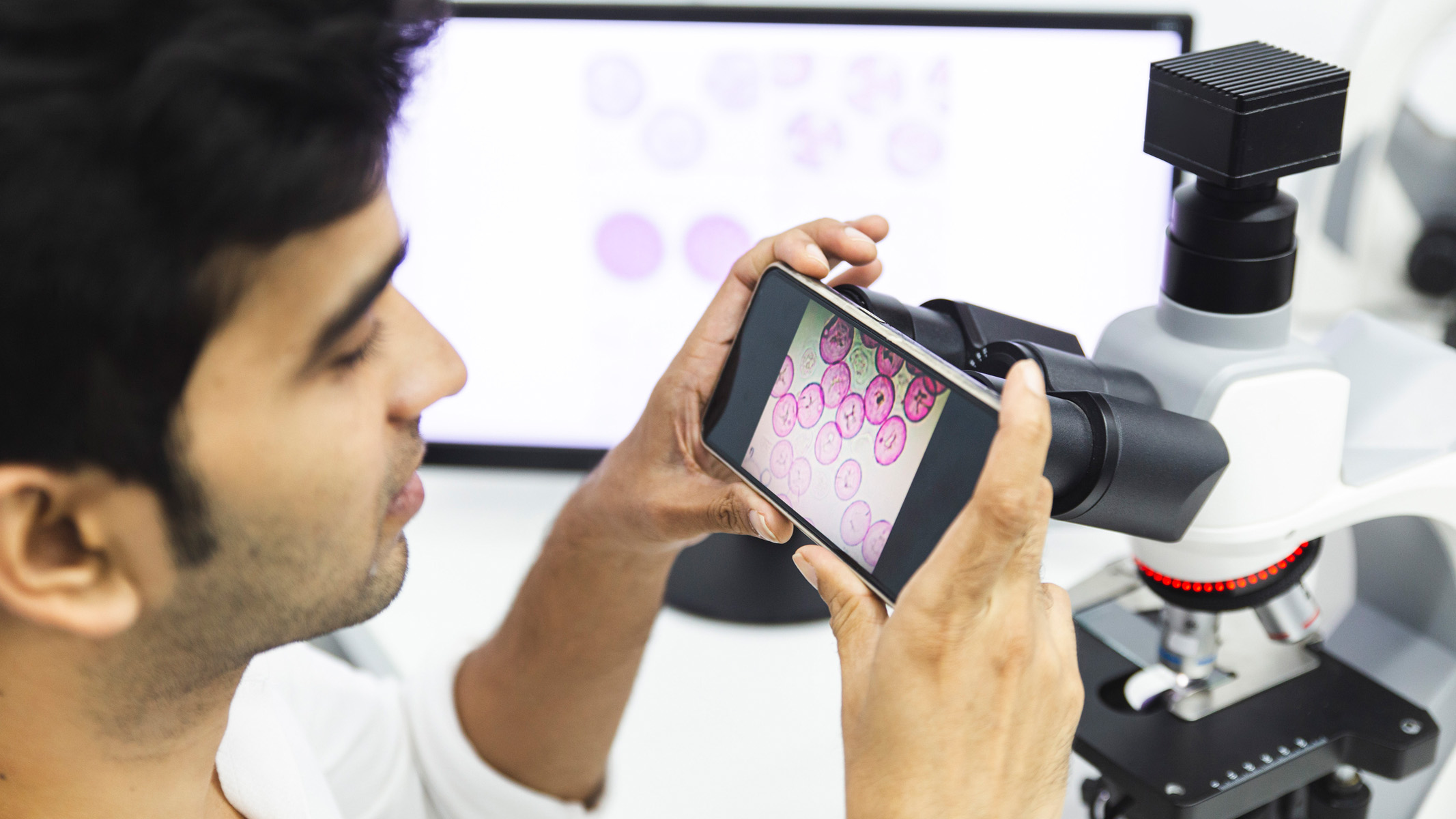
The most basic and most affordable method to take a photo of what you see through your microscopic lense is to put the electronic camera of your mobile phone or tablet over the eyepiece lens of your microscopic lense. You will require persistence and a stable hand, however it is possible to focus, focus and take photos or video in the normal method for your gadget.
Keep in mind that, before cropping, images will reveal the entire field of vision and bodytube case. To make mobile phone photomicrography much easier, you might attempt utilizing a universal smart device install, which will leave both hands totally free for running the electronic camera and decrease electronic camera shake.
For more expert outcomes and greater quality images, a one of the finest video cameras can likewise be connected to a microscopic lense. A DSLR is the apparent option for maximum resolution photomicrography, increased information at low light levels and a larger series of lens alternatives.
Get the world’s most interesting discoveries provided directly to your inbox.[
- Read our complete Swift SW200DL Compound Monocular Microscope evaluation
Photographing particular topics
When thinking about how to establish your specimens for photomicrography, the basic general rule is to utilize the exact same conditions that you would utilize for maximum watching. Attaining a clear, high-resolution image that is well contrasted, juxtaposed and in focus will provide you the very best opportunity of recording a high quality image. Photos can be digitally controlled later on, some microscopic lens come with a choice of coloured filters that sit above the condenser. These boost specific cellular structures and colours and deserve try out.
For cell photomicrography, to expose the subcellular structures within, we would advise a substance microscopic lense, with an adjustable lower light, that can amplify 400x or more. Accomplishing the proper brightness is important for sufficient direct exposure times. Varying the light is an excellent location to begin: ground glass can be utilized to diffuse light, or you might attempt utilizing a neutral density(ND) filter to minimize light strength without impacting colour.
Lots of typical spots utilized in cellular microscopy do not photo well and can provide a muddy look. Utilizing a didymium filter to obstruct orange/yellow waves brings more brilliant colours to your cell photomicrographs. Furthermore, if you discover a blue cast over the specimen background, attempt an ultraviolet light-absorbing filter. This must enhance contrast and offer a cleaner background.
- Read our complete Celestron Labs S10-60 Stereo Microscope evaluation
Photographing the endurance of ivy-leaved toadflax utilizing an old SciChem substance microscopic lense and iPad, leading and bottom brightened at 100x zoom. (Image credit: Heather Barker)
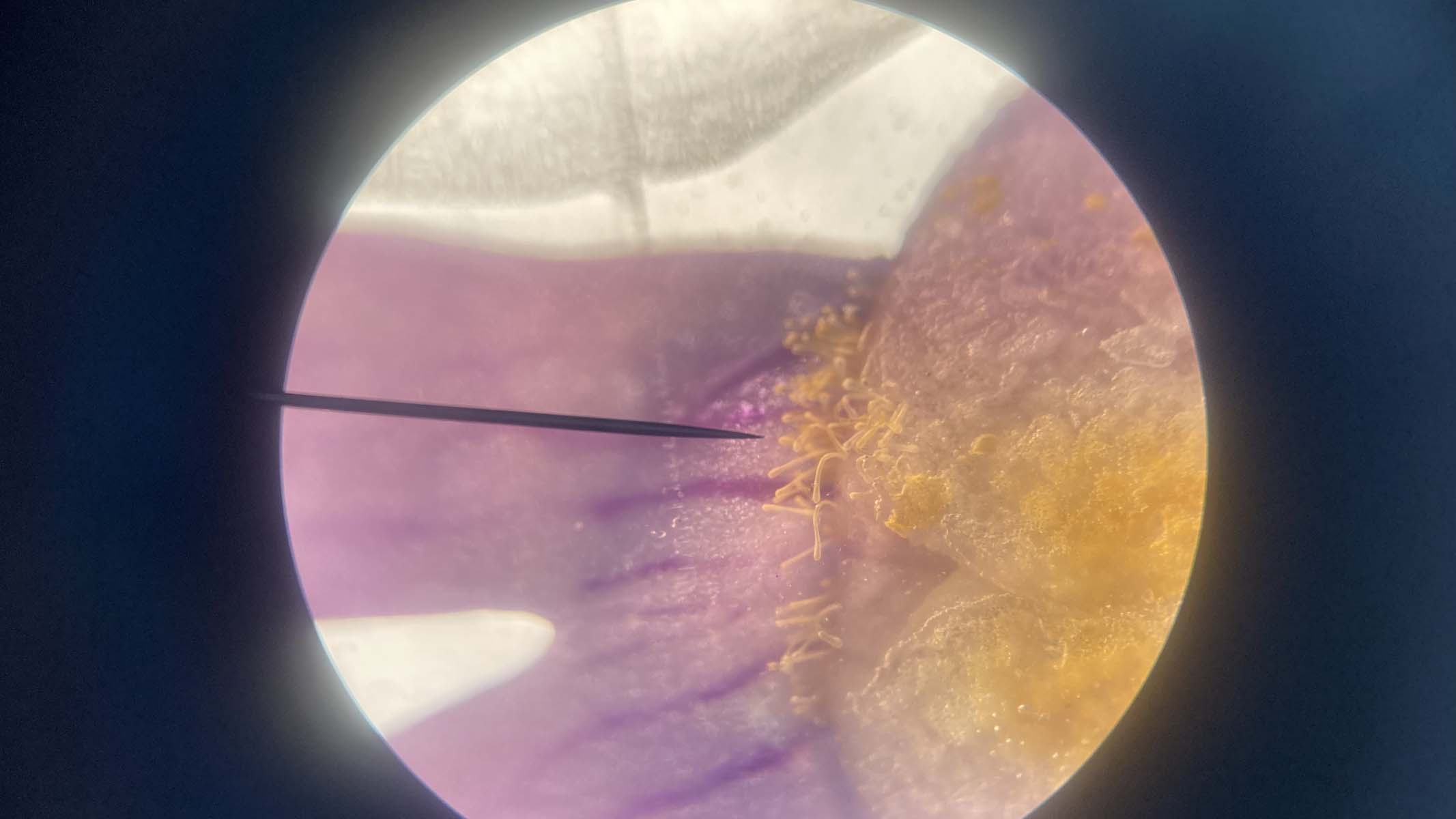
Nontransparent specimens like pollen grains or seeds can not send light, so select a microscopic lense with an upper source of light to show illuminate through the instrument. It can likewise be advantageous to establish an independent spotlight, to shine from the side and emphasize particular locations or boost colours. Some microscopic lens with upper lighting included interchangeable phase plates that are terrific for differing contrast. Black, white and frosted plates are the most typical. You might likewise attempt making your own from colored card or acetate.
Photographing single-celled organisms is comparable to cell photomicrography, as long as you are ready to eliminate and repair your specimens. Quickly discovered in water samples, tiny living organisms permit light to go through them, which exposes their internal structures. You can observe the contents of the gastrointestinal system and internal organs of small water fleas.
Establishing the school SciChem substance microscopic lense with lower lighting and side spotlight, all set for photomicrography. (Image credit: Heather Barker)
Obviously, it is far more gentle and fascinating to observe the anatomy and motion of living organisms. This is not without its difficulties! Organisms that are mobile, like tardigrades, can vanish out of view rapidly. Videoing your sample at low zoom is a wonderful method to take a look at the motion patterns of living organisms. A digital microscopic lense is indispensable for recording the tiny living world. Utilize your mobile phone install with the video function. Later on, you can pick any still images from your digital video footage.
If you are exceptionally fastidious and have a stable hand, or electronic camera install, you might take a series of successive images as you move down the focus layers and focus stack and mix them in Photoshop. This mimics depth and offers a three-dimensional look to your last image.
Heather Barker is Head of Science at All Hallows Prep-School in Somerset, England. She has actually been informing youths in between the ages of 5 and 19, as an instructor and personal tutor, considering that 2012. Studying graphics with illustration to keep herself psychologically challenged while bringing up her household, Heather re-trained by carrying out a Science degree with the Open University. Finished a Master’s degree in Developmental Biology and Biochemistry at The University of Bath in 2010. Later, Heather began work at the university as a Research Assistant, utilizing histology and genes to study the development of sticklebacks, before certifying as an instructor in the Graduate training program.
Learn more
As an Amazon Associate I earn from qualifying purchases.