
As an Amazon Associate I earn from qualifying purchases.

(Image credit: Courtesy UVA Health)
A brand-new organelle has actually been found in human cells– and researchers call it a “hemifusome.”
Like the full-size organs in our bodies, the organelles within cells are specialized structures that perform particular functions. While observing filaments that preserve the shape of cells, Seham Ebrahiman assistant teacher at the University of Virginia, and her group saw a brand-new structure that was regularly appearing in the 3D images they were making.
What in the beginning appeared like an artifact in the images ended up being a brand-new organelle that might be associated with arranging, recycling and disposing of proteins within human cells. Ebrahim compared the hemifusome’s shape to that of a snowman using a headscarf; photo a little head connected to a bigger body, with a thin border separating the 2 ends.
The organelle is around 100 nanometers in size, less than half the size of even a little mitochondrion, the well-known powerhouse of the cell.
The researchers had the ability to observe the hemifusomes due to the fact that they utilized an approach called cryo-electron tomography (cryo-ET) to create their images. This method included quickly freezing cells from 4 lab-raised cell lines to protect as much of their structures as possible, making it possible to produce clear, 3D images.
“It’s like a snapshot in time without any kind of chemical or any kind of stain,” Ebrahim informed Live Science. Utilizing this imaging method, they might look within cells in a “very native state,” as if they were glass balls, she stated.
Related: Fulfill the ‘frodosome,’ a brand name brand-new organelle
Get the world’s most interesting discoveries provided directly to your inbox.
In their paper, released in the journal Nature Communications in May, the scientists composed that the severe processing actions that other imaging methods subject cells to most likely avoided hemifusomes from being observed previously. In addition, with other strategies that utilize imaging to study the traffic unfolding in a living cell, the organelle was most likely too little to be seen, appearing at most as a blur, Ebrahim included.
Ebrahim and her associates were taking a look at a setup of blisters they had actually never ever observed before. Blisters are balloon-like structures utilized to carry things like proteins and hormonal agents within and in between cells. The brand-new research study exposed 2 blisters merged together with a two-layer barrier of fat in between them.
“Even from a biophysics perspective, it’s a breakthrough,” Ebrahim stated, “because biophysically, people have always predicted, or theorized, that vesicles can exist in this hemifused state … but this was the first time that it has actually been seen in a living cell.” This observation motivated the name hemifusome, because hemifusion describes the partial merger of 2 bilayers.
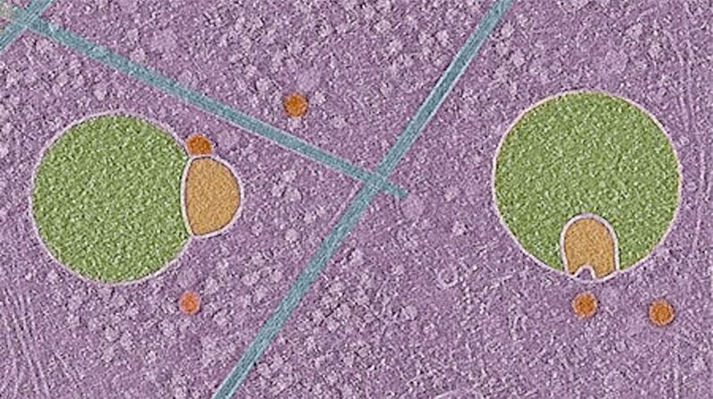
This cryo-electron tomography image( left )and 2 matching schematics(center and right)highlight the distinct structure of a hemifusome, where 2 vesicle membranes link through a hemifusion diaphragm. (Image credit: Courtesy UVA Health)
Ebrahim argues that hemifusomes can be categorized as organelles since they are self-contained practical systems within a cell, rather than “fleeting” structures that momentarily look like membranes form and divide. In the paper, she composed that it’s not likely hemifusomes are artifacts of cryo-ET.
Ebrahim’s findings “suggest that the hemifusomes they visualize are genuine cellular intermediates, not freezing-induced distortions,” stated Yi-Wei Changan assistant teacher of biochemistry and biophysics at the University of Pennsylvania Perelman School of Medicine who was not associated with the work.
When the function and function of hemifusomes have actually been validated through more research studies, they might be acknowledged as their own class of intermediate structures that meet part of blend procedures in mammalian cells, Chang informed Live Science in an e-mail.
With their existing work, the scientists can verify that the hemifusome exists, however they have yet to identify the organelle’s precise function, its life process or its structure. Ebrahim assumes that hemifusomes are precursors to particular kinds of blisters. She thinks hemifusomes might play a vital function in the recycling or disposal of cellular membranes, which is necessary for avoiding the accumulation of things in cells that might gum up their operations if permitted to build up.
The scientists composed that comprehending more about how hemifusomes work might likewise open brand-new insight into how illness such as Alzheimer’s manifest. Alzheimer’s illness is connected to the incorrect clearance of irregular protein plaques in the brain, which causes accumulation in time.
“Without cryo-electron tomography, we would have missed this discovery,” Ebrahim stated, including that “there’s probably a whole world out there that we still have to find.”
Christoph Schwaiger is an independent reporter. His primary locations of focus are science, innovation and existing affairs. His work has actually appeared in a variety of recognized outlets in different nations. When he’s not hectic hosting the conversations himself, Schwaiger is likewise a routine visitor on various news programs and programs. He likes being active and is routinely spotted assisting companies that promote causes that are close to his heart. Schwaiger holds an MA in journalism.
Learn more
As an Amazon Associate I earn from qualifying purchases.